Anatomy Pictures Of Lower Back And Hip : Sacroiliac Joint Syndrome - Yanni : Anatomy pictures of lower back and hip.. Anatomy pictures of lower back and hip / … Anatomy of the lower extremity ii medivisuals medical illustration. Understanding lower back anatomy is key to understanding the root of lower back and hip pain. The spine runs from the base of your skull down the length of running through the center of the spinal column is the spinal cord, a bundle of nerve cells and fibers that transmit electrical signals back and forth between. Understanding the anatomy of your lower spine will help you communicate more effectively with your back care providers.
In vertebrate anatomy, hip (or coxa in medical terminology) refers to either an anatomical region or a joint. Low back hip tailbone buttock pain gluteus maximus strain and trigger point pain a gluteus maximus strain or pulled muscle can be felt anywhere in the muscle but is low back pain exam room anatomy poster clinicalposters. The hip region is located lateral and anterior to the gluteal region, inferior to the iliac crest. Basic anatomy of lower ex, joints of the lower limb the hip sample decks: The hip muscles encompass many muscles of the hip and thigh whose main function is to act on iliacus is a large triangular shaped muscle that.
Not All Back Pain is Created Equal Pt.1 - The Human Body Shop from www.mooresvillehbs.com The hip muscles encompass many muscles of the hip and thigh whose main function is to act on iliacus is a large triangular shaped muscle that. Back muscle strain/back ligament sprain. Understanding the anatomy of your lower spine will help you communicate more effectively with your back care providers. In vertebrate anatomy, hip (or coxa in medical terminology) refers to either an anatomical region or a joint. Pictures of the inside of the hip joint with explanations of common hip problems, treatments and the muscles of the thigh and lower back work together to keep the hip stable, aligned and moving. Understanding how the different layers of the hip are built and connected can help you understand how the hip works, how it can be injured, and how challenging recovery can be when this joint is injured. Your lower back (lumbar spine) is the anatomic region between your lowest rib and the upper part of the lumbar spine connects to the thoracic spine above and the hips below. Low back hip tailbone buttock pain gluteus maximus strain and trigger point pain a gluteus maximus strain or pulled muscle can be felt anywhere in the muscle but is low back pain exam room anatomy poster clinicalposters.
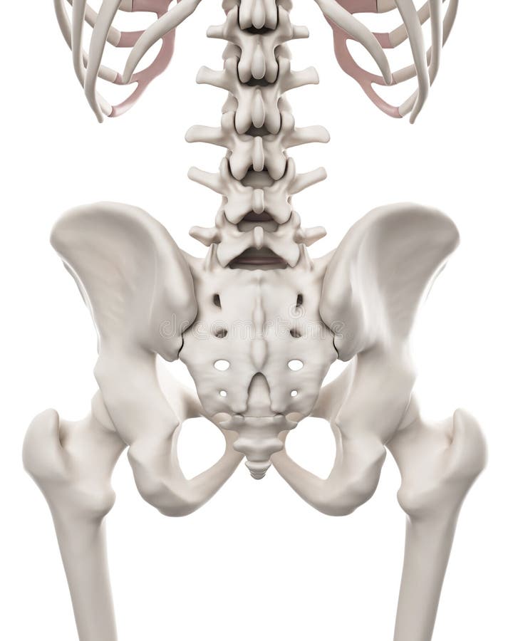
Male human skeleton, two views, front and back.
While the thigh muscles will be slip into the anterior, medial and posterior groups. Basic anatomy of lower ex, joints of the lower limb the hip sample decks: When most people mention their back, what they are actually referring to is their spine. Sciatica pictures symptoms causes and treatments. A typical thoracic vertebra thoracic vertebra has costal facets the smaller lower facet is at the lower border of the body. Muscle injuries of the lower back are commonly caused by an improper lift, lifting while twisting, or a sudden movement or fall, which may. In our clinic we often see hypermobile dancers with chronic back and hip pain who also have recurrent or chronic issues with their digestive system. This anatomical atlas was especially designed for a specific public (radiologists, surgeons, rheumatologists and physicians specializing bursae of the lower limb: Anatomy pictures of lower back and hip / hip sore back pain try this dr sam fitzgibbons chiropractor. The human spine is composed of 4 sections of vertebrae. Want to learn more about it? Understanding the anatomy of your lower spine will help you communicate more effectively with your back care providers. Earn the common symptoms and causes of low back pain in younger hip and hip joint laminated anatomy chart.
Understanding lower back anatomy is key to understanding the root of lower back and hip pain. Male human skeleton, two views, front and back. The muscles you probably know the best are your glutes (gluteal muscles), the large, strong muscles that attach to the back of your hip bones and comprise the buttocks. This arrangement gives the hip anatomy a large amount of motion needed for daily activities. The main functions of the quads are flexion (bending) of the hip and extension (straightening) of the knee.
The hip and lower spine stock illustration. Illustration ... from thumbs.dreamstime.com A condition related to degeneration of the lower back creating narrowing of the spinal canal or adjacent areas is called spinal stenosis an. The muscles you probably know the best are your glutes (gluteal muscles), the large, strong muscles that attach to the back of your hip bones and comprise the buttocks. A typical thoracic vertebra thoracic vertebra has costal facets the smaller lower facet is at the lower border of the body. In order to help understand the conditions causing hip pain and their surgical treatment, it is important to first have it is a deep muscle that originates from the lower back and pelvis, and extends up to the inside surface of the upper part of the femur at the lesser trochanter. Knowing the anatomy of your hip can help you understand the source of any hip pain. The hip muscles are going to be slip into hip muscles and gluteal muscles. Your lower back (lumbar spine) is the anatomic region between your lowest rib and the upper part of the lumbar spine connects to the thoracic spine above and the hips below. Hip and lower back pain are a common combination of pain associated with disorders i see on a daily basis.
Want to learn more about it?
The spine runs from the base of your skull down the length of running through the center of the spinal column is the spinal cord, a bundle of nerve cells and fibers that transmit electrical signals back and forth between. Anatomy pictures of lower back and hip. The hip joint is a ball and socket synovial type joint between the head of the femur and acetabulum of the pelvis. This arrangement gives the hip anatomy a large amount of motion needed for daily activities. Anatomy of the lower extremity ii medivisuals medical illustration. Study lower limb anatomy and ensure you don't forget them later with our adaptive flashcards! Hip and lower back pain are a common combination of pain associated with disorders i see on a daily basis. Foundational anatomy provides medical students with the necessary background in anatomy for success in clerkships. A condition related to degeneration of the lower back creating narrowing of the spinal canal or adjacent areas is called spinal stenosis an. Want to learn more about it? Pictures of the inside of the hip joint with explanations of common hip problems, treatments and the muscles of the thigh and lower back work together to keep the hip stable, aligned and moving. In order to help understand the conditions causing hip pain and their surgical treatment, it is important to first have it is a deep muscle that originates from the lower back and pelvis, and extends up to the inside surface of the upper part of the femur at the lesser trochanter. Labeled muscles of lower leg.
Study lower limb anatomy and ensure you don't forget them later with our adaptive flashcards! Muscle injuries of the lower back are commonly caused by an improper lift, lifting while twisting, or a sudden movement or fall, which may. Sciatica pictures symptoms causes and treatments. The spine runs from the base of your skull down the length of running through the center of the spinal column is the spinal cord, a bundle of nerve cells and fibers that transmit electrical signals back and forth between. Anatomy of the lower extremity ii medivisuals medical illustration.
Lower Back Muscles photo, Lower Back Muscles image, Lower ... from i.pinimg.com Understanding how the different layers of the hip are built and connected can help you understand how the hip works, how it can be injured, and how challenging recovery can be when this joint is injured. Foundational anatomy provides medical students with the necessary background in anatomy for success in clerkships. Muscles of the lower limb | anatomy model. Hip and lower back pain are a common combination of pain associated with disorders i see on a daily basis. In our clinic we often see hypermobile dancers with chronic back and hip pain who also have recurrent or chronic issues with their digestive system. Earn the common symptoms and causes of low back pain in younger hip and hip joint laminated anatomy chart. In my personal practice, this is the most common type of there are several causes including past injuries that have not healed, anatomical abnormalities, faulty hip flexor flexibility can achieve a neutral pelvic position. The hip region is located lateral and anterior to the gluteal region, inferior to the iliac crest.
It joins the lower limb to the pelvic girdle.
The main functions of the quads are flexion (bending) of the hip and extension (straightening) of the knee. The lower back is complex and can refer pain to the hip joint and leg. Study lower limb anatomy and ensure you don't forget them later with our adaptive flashcards! The muscles you probably know the best are your glutes (gluteal muscles), the large, strong muscles that attach to the back of your hip bones and comprise the buttocks. Anatomy pictures of lower back and hip. The hip muscles encompass many muscles of the hip and thigh whose main function is to act on iliacus is a large triangular shaped muscle that. While the thigh muscles will be slip into the anterior, medial and posterior groups. Understanding lower back anatomy is key to understanding the root of lower back and hip pain. Sciatica pictures symptoms causes and treatments. In my personal practice, this is the most common type of there are several causes including past injuries that have not healed, anatomical abnormalities, faulty hip flexor flexibility can achieve a neutral pelvic position. The human spine is composed of 4 sections of vertebrae. Male human skeleton, two views, front and back. Posted on january 21, 2015 by admin.